
A skin imaging system is a clinical technology designed to capture, enlarge, and analyze visible skin features. It may use high-resolution cameras, polarized light, ultraviolet illumination, or 3D scanning. These tools can reveal details that ordinary room lighting often hides. A small brown spot, uneven border, or subtle texture change may become clearer on a calibrated screen. The system does not simply take photographs. It creates standardized images that support careful comparison over time.
In dermatology clinics, trained professionals may use these images during examinations, treatment planning, and follow-up visits. Some systems measure color, lesion size, vascular patterns, or changes in skin texture. Software can assist with documentation and image organization. However, it should support clinical judgment rather than replace it. The quality of results depends on lighting, camera settings, patient positioning, and operator experience. Small differences matter.
Not every visible change indicates disease. Skin can look different after sun exposure, inflammation, medication, or minor injury. This is where reliable interpretation becomes important. A qualified clinician considers the image alongside medical history, physical examination, and, when appropriate, further testing. The technology has limits. It can miss findings, produce false alerts, or encourage overconfidence when used without context. Understanding how a skin imaging system works helps patients and professionals use its strengths responsibly, while recognizing what the image cannot prove.
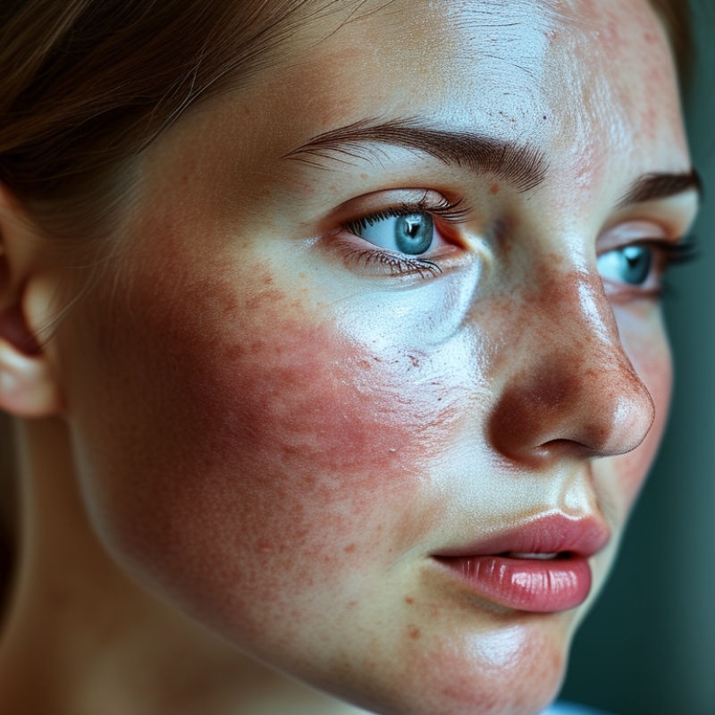
A skin imaging system is a clinical tool that captures, enlarges, and compares images of the skin. It may use standard photography, dermoscopy, polarized light, or three-dimensional scanning. Some systems also apply software to measure color, border shape, lesion size, and visual changes over time. A trained clinician reviews these findings. The technology supports examination; it does not replace professional judgment.
The value becomes clearer during monitoring. A 2022 International Agency for Research on Cancer report recorded about 332,000 new melanoma cases worldwide. Small visual changes can therefore deserve careful attention. During an appointment, a clinician may photograph a mole under controlled lighting, record its location, and compare it with earlier images. Some systems create a body map, helping identify new or changing spots that ordinary memory may miss. Useful detail matters.
Accuracy still has limits. Image quality can fall with hair, shadows, movement, or inconsistent camera distance. Artificial intelligence may highlight suspicious patterns, but it can also produce false positives or overlook unusual lesions. A 2021 systematic review in the British Journal of Dermatology found that AI performance varied across studies, devices, and patient groups. That finding deserves reflection. Skin tone representation also remains uneven in medical datasets, which may affect reliability. Results should be interpreted with medical history, physical examination, and, when necessary, laboratory testing. A skin imaging system is best understood as structured visual evidence, not an automatic answer.
A skin imaging system combines specialized cameras, controlled lighting, software, and clinical records. It captures surface details that ordinary room lighting can hide. It is not a magic camera. Its value depends on consistent images and trained interpretation.
The imaging unit usually includes a high-resolution camera, polarized light, and close-up lenses. Polarized illumination reduces glare, while cross-polarized images can reveal redness and pigment patterns. Some systems add ultraviolet light, though this feature requires careful clinical validation. A dermoscopic lens provides magnified views of structures beneath the skin surface. Measurement tools record lesion diameter, border changes, and location. Software then stores images, compares visits, and flags visible differences. However, automated alerts are not diagnoses.
Reliable systems also need color calibration, steady positioning, and even exposure. A small shift in camera angle can make a mole appear wider. This is easy to overlook. Secure data storage matters too, because images are linked to sensitive health information. The International Agency for Research on Cancer recorded about 332,000 new melanoma cases worldwide in 2022, showing why dependable monitoring matters (IARC Global Cancer Observatory, 2024). The American Academy of Dermatology reports that approximately one in five Americans will develop skin cancer by age 70. These figures support regular professional assessment, but imaging alone cannot replace it. A clear image may still miss texture, symptoms, or changes outside the captured frame.
A skin imaging system captures detailed photographs under controlled conditions. It usually combines a high-resolution camera, fixed lighting, and positioning guides. The patient rests the chin and forehead against supports. This reduces movement and keeps the viewing angle consistent.
The system may photograph the face from several directions. Front, left, right, and close-up views reveal different surface details. Some devices use polarized light to reduce glare from oil and moisture. Others add ultraviolet illumination to show certain pigmentation patterns beneath the surface. Before capture, the operator checks focus, exposure, and skin cleanliness. Even a loose strand of hair can hide a small area.
Consistent lighting matters greatly. Brightness, color temperature, and camera distance should remain stable during repeat visits. Specialized software then compares images and maps features such as spots, redness, pores, and wrinkles. It can measure changes, but it does not replace professional clinical judgment. A camera records appearance, not the complete biological cause.
In practice, results depend on the operator. Facial expression, recent exercise, or skincare products may affect the image. The process is not flawless. I would treat automated scores as supporting evidence, not final truth. A qualified professional should review unusual findings, especially when an area changes in shape, color, or texture. Clear records also help patients understand what the images can—and cannot—show.
| System Dimension | How It Works | Skin Information Captured | Typical Output | Important Considerations |
|---|---|---|---|---|
| Controlled Illumination | The system uses a consistent light source and controlled positioning to reduce changes caused by room lighting, shadows, and viewing angle. | Visible surface features such as color variation, redness, pigmentation, dryness, texture, and apparent blemishes. | Standardized photographs that can be compared over time or between different areas of the skin. | Brightness, color temperature, camera distance, and subject positioning should remain consistent for reliable comparisons. |
| Visible-Light Imaging | A digital camera records reflected light from the skin in the visible portion of the electromagnetic spectrum. | Surface color, lesions, spots, wrinkles, pores, irritation, and changes in skin texture. | Color images that document the visible appearance of the skin. | Images show appearance rather than providing a definitive medical diagnosis. Clinical interpretation may be required. |
| Cross-Polarized Imaging | Polarizing filters are arranged to reduce surface glare, allowing the camera to record more of the light scattered from beneath the skin surface. | Features that may be less visible in ordinary lighting, including some redness, pigmentation patterns, and subsurface color variation. | Images with reduced specular reflection and improved visibility of selected subsurface features. | Results depend on filter orientation, lighting geometry, skin type, and image-processing methods. |
| Ultraviolet Imaging | The system illuminates the skin with ultraviolet light and records differences in the way skin areas absorb, reflect, or fluoresce under that illumination. | Some surface oil patterns, pigment-related contrast, and areas of unevenness that may not be obvious in visible light. | Contrast-enhanced images that highlight selected optical differences on the skin surface. | Ultraviolet images are not equivalent to visible-light photographs and should be interpreted cautiously. Eye and skin safety controls are essential. |
| Multispectral Imaging | The system captures images in multiple defined wavelength bands rather than relying on a single broad color image. | Wavelength-dependent differences related to pigmentation, redness, blood-related optical properties, and surface condition. | A set of aligned images or calculated maps showing changes across selected spectral bands. | Measurements can be affected by ambient light, calibration quality, motion, and differences in camera sensitivity. |
| Image Capture and Focus | A lens projects light onto an image sensor, while autofocus or fixed-focus settings help maintain sharpness across repeated captures. | Fine details such as lines, scales, pore appearance, edges of spots, and localized texture changes. | High-resolution digital images suitable for visual review, measurement, or automated analysis. | Focus, motion blur, lens cleanliness, and image compression can reduce the visibility of small features. |
| Positioning and Distance Control | A headrest, face guide, measurement scale, or fixed camera position helps keep the subject at a repeatable distance and angle. | Comparable images of the same region during follow-up examinations or skincare assessments. | Consistent image framing and, when calibrated, approximate spatial measurements. | Changes in pose, facial expression, camera angle, or distance can create apparent differences that are not caused by the skin itself. |
| Color Calibration | Reference targets or calibration procedures are used to compensate for variations in illumination and camera response. | More consistent representation of skin tone, redness, pigmentation, and color-based changes. | Images with improved color consistency across sessions and imaging conditions. | Calibration improves comparability but does not eliminate all effects from ambient conditions, cosmetics, tanning, or physiological changes. |
| Three-Dimensional Surface Imaging | Multiple images or projected patterns are combined to estimate the shape and depth of the skin surface. | Contours, depressions, raised areas, wrinkles, scars, and changes in surface relief. | A three-dimensional model, depth map, or surface-height measurement. | Hair, movement, reflective products, and complex geometry may interfere with accurate surface reconstruction. |
| Image Processing | Software may correct illumination, align images, remove background areas, enhance contrast, and calculate selected image features. | Quantifiable characteristics such as color values, area, intensity, texture, contrast, or lesion dimensions. | Annotated images, measurement tables, heat maps, trend graphs, or standardized scores. | Processing can improve consistency but may also introduce artifacts if the algorithm or settings are unsuitable. |
| Longitudinal Comparison | Images captured at different times are aligned and compared to identify changes in the same skin region. | Trends in visible pigmentation, redness, lesion size, wrinkles, texture, or other documented features. | Side-by-side comparisons, difference maps, and time-based progress reports. | Comparisons are most meaningful when lighting, positioning, camera settings, skincare products, and environmental conditions are controlled. |
| Data Storage and Privacy | Images and associated measurements are stored with identifiers, timestamps, and examination details for later retrieval and comparison. | The image record, capture conditions, analyzed regions, and any user-entered observations. | Searchable image files, measurement records, and secured reports. | Facial and skin images can be sensitive personal data. Access control, secure storage, consent, and appropriate retention policies are important. |
A skin imaging system converts a visible skin surface into measurable data.
Its camera captures several images under controlled lighting, distance, and focus. Some systems also use polarized or multispectral light. This can reveal redness, pigmentation, texture, and vascular patterns that ordinary photographs may miss.
Software then corrects color balance and removes shadows. It separates the lesion from surrounding skin through image segmentation. Next, algorithms measure diameter, asymmetry, border irregularity, color variation, and surface texture. Machine-learning models compare these features with trained image databases. The output may show risk scores, change maps, or side-by-side progress images. It does not replace clinical judgment. The World Health Organization and International Agency for Research on Cancer estimated 1.5 million non-melanoma skin cancer cases and 330,000 melanoma cases worldwide in 2022. Reliable analysis therefore needs careful follow-up, not a single automated result. Image quality still matters greatly.
Tips: Capture images in the same room, distance, and lighting. Keep the lens clean. Record the body location and date. Ask whether the system was tested across different skin tones. This point is often overlooked. A polished image can still produce an uncertain result. Studies published in Nature Medicine have shown that artificial intelligence can support dermatologists, but performance depends on data quality and clinical context. WHO data also reminds us that large populations are affected, while local validation remains essential.
What Is a Skin Imaging System and How Does It Work?
A skin imaging system captures detailed photographs of the skin using controlled lighting, magnification, polarized filters, or multiple wavelengths. Some systems also create three-dimensional surface maps. Software then measures features such as redness, pigmentation, pores, wrinkles, texture, and lesion borders. A trained professional compares images over time rather than relying on one snapshot. The process sounds objective. It is not completely automatic.
Applications and Limitations of Skin Imaging Systems
Clinics use these systems to document a patient’s baseline condition and monitor visible changes during treatment. They can support assessments of acne, uneven pigmentation, sun-related damage, scars, and suspicious lesions. In research, standardized images help investigators compare treatment responses more consistently. They may also improve communication because patients can see subtle changes on a magnified screen.
However, imaging cannot replace clinical examination, medical history, or tissue testing when needed. Lighting, camera distance, skin moisture, makeup, and movement can alter results. Dark hair may hide a lesion. Shadows may create one. Algorithms can also perform unevenly across different skin tones if their training data lacks diversity. This limitation deserves more attention. A high score is not a diagnosis, and a normal-looking image does not guarantee healthy skin. Reliable use requires calibrated equipment, consistent image capture, trained reviewers, informed consent, and secure data handling. Even experienced clinicians may disagree when changes are slight.
"*" indicates required fields